CBSE Class 12 Human Reproduction class 12 Notes Biology in PDF are available for free download in myCBSEguide mobile app. The best app for CBSE students now provides Human Reproduction class 12 Notes latest chapter wise notes for quick preparation of CBSE board exams and school-based annual examinations. Class 12 Biology notes on chapter 3 Human Reproduction are also available for download in CBSE Guide website.
CBSE Guide Human Reproduction class 12 Notes Biology
CBSE guide notes are the comprehensive notes which covers the latest syllabus of CBSE and NCERT. It includes all the topics given in NCERT class 12 Biology textbook. Users can download CBSE guide quick revision notes from myCBSEguide mobile app and my CBSE guide website.
Class 12 Biology notes Chapter 3 Human Reproduction
Download CBSE class 12th revision notes for chapter 3 Human Reproduction in PDF format for free. Download revision notes for Human Reproduction class 12 Notes and score high in exams. These are the Human Reproduction class 12 Notes prepared by team of expert teachers. The revision notes help you revise the whole chapter 3 in minutes. Revision notes in exam days is one of the best tips recommended by teachers during exam days.
Download Revision Notes as PDF
CBSE Class 12 Biology Revision Notes Chapter 3 Human Reproduction
Humans are sexually reproducing and viviparous. The reproductive events in humans include formation of gametes (gametogenesis), i.e., sperms in males and ovum in females, transfer of sperms into the female genital tract (insemination) and fusion of male and female gametes (fertilisation) leading to formation of zygote. This is followed by formation and development of blastocyst and its attachment to the uterine wall (implantation), embryonic development (gestation) and delivery of the baby (parturition)
The Male Reproductive System: It cChapteronsists of:
a) Primary sex organs i.e. a pair of testes suspended in a scrotum.
b) Secondary sex organs i.e. a pair of ducts each differentiated into rete testis, vasa efferentia, epididymis and vas deferens, ejaculatory duct and the associated glands
c) External genitalia
- The testes are situated outside the abdominal cavity in a pouch called scrotum, which help in maintaining the low temperature of testes necessary for spermatogenesis.
- Each testes has about 250 testicular lobules and each lobule contain highly coiled seminiferous tubules in which sperms are produced. Each seminiferous tubules is lined by two types of cells, spermatogonia ( male germ cell) and Sertoli cells.
- Leydig cells or interstitial cells present around the seminiferous tubules synthesize and secrete androgen hormone.
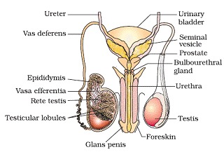
- Ejaculatory duct store and transport the sperm from testes to outside through urethra which originate from urinary bladder and extend through penis to its external opening urethral meatus.
- The penis is male external genitalia. The enlarged end of penis is called the glans penis is covered by a loose fold of skin called foreskin.
- Male accessary glands include paired seminal vesicles, prostrate and paired bulbourethral glands. Secretion of these glands forms the seminal plasma which contains fructose, calcium and enzymes. The secretion of bulbourethral glands also helps in lubrication of the penis.
The Female Reproductive System: It consists of :
a)The primary sex organ that is a pair of ovaries
b)Secondary sex organs- the duct system consisting of a pair of fallopian tube , a uterus , cervix and vagina
c)External genitalia
d)Mammary glands
- Ovaries are primary female sex organ that produce the female gamete and several steroid hormones. Each ovary is covered by thin epithelium which encloses the ovarian stroma, which is divided into a peripheral cortex and an inner medulla.
- Fallopian tube extends from periphery of ovary to the uterus. The part closer to ovary is a funnel shaped structure called infundibulum having finger like projection called fimbriae.
- Infundibulum leads to ampulla and join with uterus with isthmus. Uterus is pear shaped structure also called womb.
- Uterus open vagina through a narrow cervix. The cavity of cervix (cervical canal) along with vagina forms the birth canal.
- The wall of uterus has three layers of tissue:
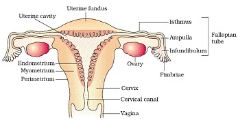
I. Perimetrium- external membrane.
II. Myometrium – middle thick layer of smooth muscles which exhibit strong contraction during delivery of baby.
III. Endometrium – line the uterine wall and undergo cyclic changes during menstrual cycle.
Female external genitalia includes
- Mons pubis – cushion of fatty tissues covered by skin and pubic hair.
- Labia majora- fleshy fold that surround the vaginal opening.
- Labia manora – paired fold of tissue under labia majora.
- The opening of vagina is often partially covered by a membrane called hymen. The tiny finger like projection present at the upper junction of two labia manora above the urethral opening is called clitoris.
Mammary glands are paired structures that contain glandular tissues and variable fats. Each glandular tissue contains 15-20 mammary lobes containing alveoli that secrete milk. Mammary ducts join to form mammary ampulla.
Gametogenesis: The process of formation of male and female gametes in testes and ovary respectively is called gametogenesis.It is of two types:
1. Spermatogenesis in males
2. Oogenesis in females
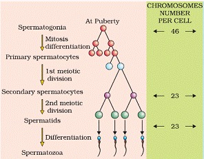
Spermatogenesis- in testes immature, male germ cells (spermatogonia) produce sperm by spermatogenesis that begin at puberty.
- The spermatogonia present at the inner side of seminiferous tubules multiply by mitotic division and increase in number. Each spematogonium contain 46 chromosomes.
- Spermatogonia forms spermatocyte that undergo meiotic division to reproduce secondary spermatocytes having 23 chromosomes.
- The spermatids are transformed into spermatozoa by the process called spermiogenesis. The sperm heads remain embedded in sertoli cells and are released from seminiferous tubules by the process of spermiation.
Hormonal control of spermatogenesis
- Spermatogenesis initiated due to increase in secretion of gonadotropin releasing hormone by hypothalamus
- Increase in GnRH act on anterior pituitary and stimulate secretion of two gonadotropins, LH and FSH
- LH acts on Leydig cells and stimulates them to secrete androgens.
- FSH acts on Sertoli cells, stimulates secretion of some factors which help in spermiogenesis
Structure of sperm- sperm is a microscopic structure composed of a head, neck, a middle piece and a tail. The sperm head contain elongated haploid nucleus, anterior portion of which is covered by cap like structure acrosome.
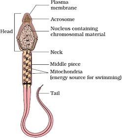
Human male ejaculates about 200-300 million sperms during a coitus. The seminal plasma along with the sperms constitutes the semen. The function of male sex secondary ducts and glands are maintained by androgen hormones.
Oogenesis : The process of formation of mature female gametes is called oogenesis. It started during embryonic development stage when millions of ogonia (gamete mother cells) are formed in each fetal ovary.
- The gametes mother cells start division and enter into prophase-I of meiotic division and get temporally arrested at that stage called primary oocytes.
- Each primary oocyteget surrounded by a layer of granulosa cell than it is called the primary follicle.
- At puberty, about 60,000- 80,000 primary follicles are left in each ovary.
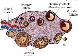
- Primary follicle gets surrounded by more layers of granulosa cells called secondary follicle that transform into tertiary follicle that contain fluid filled cavity called antrum.
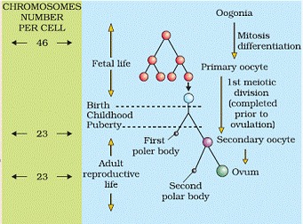
- The tertiary follicles further changes into the mature follicle called Graafian follicle, which rapture to release secondary oocytes (ovum) from the ovary by the process of ovulation.
Menstrual cycle: The reproductive cycles in female primates is called menstrual cycle. It start at puberty and is called menarche.
Phases of Menstrual Cycle
The menstrual cycle consists of following four phases:
(1) Menstrual Phase:
(i) In a 28 days menstrual cycle,the menses takes place on cycle days 3-5.
(ii) The production of LH from the anterior lobe of the pituitary gland is reduced.
(iii) The withdrawal of this hormone causes degeneration of the corpus luteum and, therefore progestrone production is reduced.
(iv) Production of oestrogen is also reduced in this phase.
(v) The endometrium of uterus breaks down & menstruation begins.
(vi) The cells of endometrium secretions, blood & unfertilised ovum constitutes the menstrual flow.
(2) Follicular Phase:
(i) This phase usually includes cycle days 6-13 or 14 in a 28 days cycle.
(ii) The follicle stimulating hormone (FSH) secreted by the anterior lobe of the pituitary gland stimulates the ovarian follicle to secrete oestrogens.
(iii) Oestrogen stimulates the proliferation of the endometrium of the uterine wall.
(iv) The endometrium becomes thicker by rapid cell multiplication and this is accompanied by an increase in uterine glands & blood vessels.
(3) Ovulatory Phase:
(i) Both LH & FSH attain a peak level in the middle of cycle (about 14th day).
(ii) Oestrogen concentration in blood increases.
(iii) Rapid secretion of LH induces rupturing of graffian follicle and thereby the release of ovum.
(iv) In fact LH causes ovulation.
(4) Luteal Phase:
(i) Includes cycle days 15 to 28.
(ii) Corpus luteum secretes progestrone.
(iii) Endometrium thickens.
(iv) Uterine glands become secretory.
Hormonal Control of MC
(i) FSH stimulates the ovarian follicles to produce oestrogens.
(ii) LH stimulates corpus luteum to secrete progestrone.
(iii) Menstrual phase is caused by the increased production of oestrogens.
(iv) LH causes ovulation
(v) Proliferative phase is caused by the increased production of oestrogens.
(vi) Secretory phase is caused by increased production of progestrone.
Fertilisation and Implantation
The process of fusion of sperm with ovum is called fertilisation.
- During coitus (copulation) semen is released into vagina. The motile sperms swim rapidly to reach the junction of isthmus and ampulla of fallopian tube. The ovum also reaches there and fusion of gametes takes place in at ampullary-isthmic junction.
- In this acrosome of sperm undergoes acrosomal reaction and releases certain sperm lysins which dissolve the egg envelopes locally and make the path for the penetration of sperm.
- These sperm lysins contain a lysing enzyme hyaluronidase which dissolves the hyaluronic acid polymers in the intercellular spaces which holds the granulosa cells of corona radiata together; corona penetrating enzyme (that dissolves the corona radiata) and acrosin (which dissolves the zona pellucida). Then it dissolves the zona pellucida.
Cortical reaction:
(a) Immediately after the entry of a sperm into the egg, the later shows a cortical reaction to check the entry of more sperms.
(b) In this reaction, the cortical granules present beneath the egg’s plasma membrane release chemical substance between the ooplasm and the plasma membrane (vitelline membrane).
(c) These substances raise the vitelline membrane above the egg surface. The elevated vitelline membrane is called fertilization membrane.
(d) The increased space between the ooplasm and the fertilization membrane and the chemical present in it effectively check the entry of other sperm.
(e) If polyspermy occurs, that is more than one sperm enter the secondary oocyte, the resulting cell has too much genetic material to develop normally
- The haploid gametes fuse together to form diploid zygote. As the zygote moves towards the uterus, the mitotic division starts and form cleavage to change into 2, 4,8,16 celled blastomeres.
- The blastomeres with 8 to 16 cells are called morula. Morula divide to change into blastocysts .The blastomeres in the blastocyst are arranged into an outer layer called trophoblast and an inner group of cells attached to trophoblast called the inner cell mass.The outer layer of blastocyst is called trophoblast that attach with endometrium of uterus, called implantation that leads to pregnancy.
Pregnancy and embryonic development
The finger-like projections on trophoblaste after implantation called is called chronic villi that along with uterine wall forms functional unit between developing embryo and maternal body called placenta. Placenta is attached with fetus with an umbilical cord that transport food and oxygen to embryo.
- Hormones hCG (human chorionic gonadotropin), hPL (human placental lactogen) and relaxin are produced in woman only during pregnancy by placenta.
- After implantation, the inner cell mass (embryo) differentiates into an outer layer called ectoderm and an inner layer called endoderm. A mesoderm soon appears between the ectoderm and the endoderm. These three layers give rise to all tissues (organs) in adults. It is important to note that the inner cell mass contains certain cells called stem cells which have the potency to give rise to all the tissues and organs
- In human, after one month of pregnancy the embryo’s heart is formed. By the end of 2nd month limbs and digits are formed. By the end of 12 months, major organs and external genital organs are well developed. The first movement of foetus is observed in 5 months. By the end of 24 weeks body is covered with fine hair, eye lids and eyeless are formed. At the end of 9 months fetus is fully developed.
PARTURITION AND LACTATION
Parturition-the process of delivery of fully developed foetus is called parturition.
- Signals for parturition originate from the fully developed fetus and placenta inducing mild uterine contractions called Foetal ejection reflex
- It triggers the release of oxytocin from maternal pituitary
The mammary glands of female, start producing milk, to the end of pregnancy by the process of lactation. The milk produced during the initial few days of lactation is called colostrum, which contain several antibodies.
CBSE Class 12 Revision Notes and Key Points
Human Reproduction class 12 Notes Biology. CBSE quick revision note for class-12 Chemistry Physics Math’s, Biology and other subject are very helpful to revise the whole syllabus during exam days. The revision notes covers all important formulas and concepts given in the chapter. Even if you wish to have an overview of a chapter, quick revision notes are here to do if for you. These notes will certainly save your time during stressful exam days.
- Physics
- Chemistry
- Mathematics
- Biology
- Accountancy
- Economics
- Business Studies
- Computer Science
- Informatics Practices
- English Core
- History
- Physical Education
To download Human Reproduction class 12 Notes Biology, sample paper for class 12 Physics, Chemistry, Biology, History, Political Science, Economics, Geography, Computer Science, Home Science, Accountancy, Business Studies and Home Science; do check myCBSEguide app or website. myCBSEguide provides sample papers with solution, test papers for chapter-wise practice, NCERT Human Reproduction, NCERT Exemplar Human Reproduction, quick revision notes for ready reference, CBSE guess papers and CBSE important question papers. Sample Paper all are made available through the best app for CBSE students and myCBSEguide website.
- Reproduction in Organisms class 12 Notes Biology
- Sexual Reproduction in Flowering Plants class 12 Notes Biology
- Reproductive Health class 12 Notes Biology
- Principles of Inheritance and Variation class 12 Notes Biology
- Molecular Basis of Inheritance class 12 Notes Biology
- Evolution class 12 Notes Biology
- Human Health and Disease class 12 Notes Biology
- Strategies for Enhancement in Food Production class 12 Notes Biology
- Microbes in Human Welfare class 12 Notes Biology
- Biotechnology Principles and Processes class 12 Notes Biology
- Biotechnology and its Applications class 12 Notes Biology
- Organisms and Populations class 12 Notes Biology
- Ecosystem class 12 Notes Biology
- Biodiversity and Conservation class 12 Notes Biology
- Environmental Issues class 12 Notes Biology
Test Generator
Create question paper PDF and online tests with your own name & logo in minutes.
Create Now
Learn8 App
Practice unlimited questions for Entrance tests & government job exams at ₹99 only
Install Now